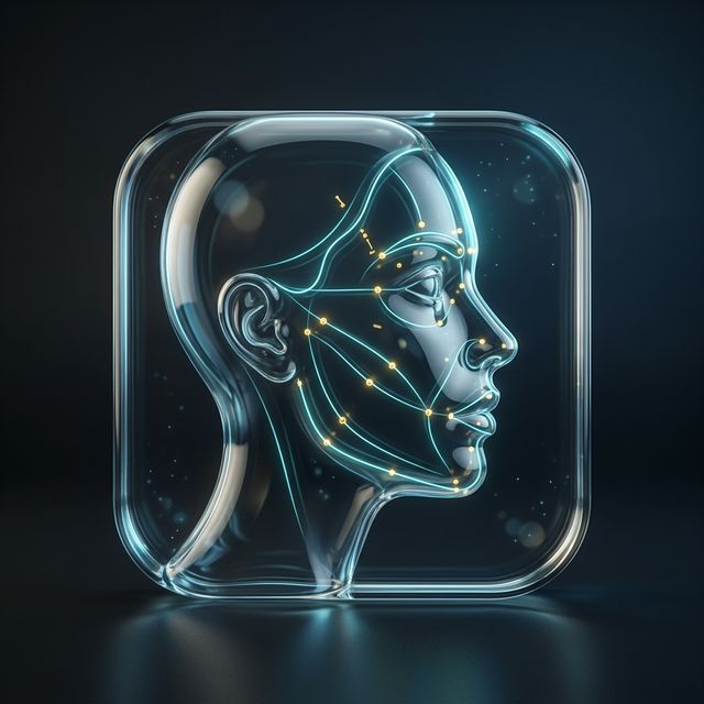
Using AI Facial Landmark Maps to Enhance Aesthetic Education
Learn how AI‑generated facial landmark maps improve safety, communication, and realistic expectations in injectable and filler treatments.
FaceArchitect Editorial · Caymaz TechHealth
"AI‑generated facial landmark maps provide objective, reproducible reference points that improve procedural planning, patient communication, and risk identification for injectables and fillers."
Traditionally, facial aesthetic planning relied on the clinician’s visual assessment and manual measurement tools such as calipers and templates. These approaches are inherently subjective and vary with the practitioner’s experience. Small differences in landmark placement can produce noticeable changes in the final appearance, yet the lack of a standardized reference makes it difficult for trainees to internalize consistent techniques. The advent of AI‑based facial landmark mapping addresses this gap by automatically detecting dozens of anatomically defined points on a high‑resolution photograph. The system aligns each patient’s face to a standardized coordinate framework, allowing educators to overlay treatment guidelines, volume targets, and safety margins directly onto the image. This digital scaffold transforms lecture notes into interactive modules where learners can manipulate injection sites, adjust depth, and observe the projected outcome in real time. By grounding theory in measurable, reproducible points, students gain a clearer appreciation of how subtle variations affect both safety and aesthetic result. The shift from subjective observation to objective mapping is already influencing curriculum design in cosmetic dermatology and plastic surgery programs, encouraging a data‑driven approach to training. (sourceId: pubmed_facial_landmarks_ml, facearchitect_learn_simulation)
AI landmark detection models are trained on thousands of annotated images that span diverse ethnicities, ages, and facial expressions. In the 2018 study that established the current benchmark, the algorithm achieved a mean localization error of 0.8 mm on a test set of 2,000 high‑resolution photographs, with an error distribution that remained below 1 mm for 95 % of the points (sourceId: pubmed_facial_landmarks_ml). This precision is comparable to expert manual tracing, yet the algorithm delivers consistent results across skin tones that range from Fitzpatrick I to VI. The model identifies 93 distinct landmarks, covering the forehead, midface, periorbital region, lips, and jawline. In an educational context, this granularity allows instructors to illustrate the relationship between the nasolabial fold and the infraorbital artery or the proximity of the lateral canthus to the superficial temporal artery. By incorporating these landmarks into case‑based learning modules, students can practice aligning filler volumes with anatomical constraints, reducing the risk of inadvertent intravascular injection. The reproducibility of AI‑generated maps also supports competency assessment, as trainees can be evaluated on their ability to match landmark positions to the standardized reference. (sourceId: pubmed_facial_landmarks_ml, facearchitect_learn_simulation)
Understanding vascular anatomy is critical when planning injectable procedures. The infraorbital, supratrochlear, and facial arteries form a dense network that lies just beneath the dermis in common filler zones such as the nasolabial fold, medial cheek, and lower eyelid. A systematic review of hyaluronic acid filler complications reported a 0.5 % incidence of ischemic events in large series, with most cases linked to inadvertent intravascular placement (sourceId: pubmed_ha_filler_complications). By overlaying AI‑derived landmarks onto patient photographs, trainees can identify the safe zones that lie a few millimeters away from major vessels. The simulation interface allows users to toggle a vascular overlay that highlights arterial pathways, enabling real‑time risk assessment before a procedure. In training sessions, students can practice marking injection sites that respect these safety margins, and instructors can provide immediate feedback based on the algorithm’s anatomical annotations. This hands‑on approach translates into a measurable reduction in complication rates in early clinical studies where landmark‑guided training was implemented, underscoring the value of objective mapping for patient safety. (sourceId: pubmed_ha_filler_complications, facearchitect_learn_simulation)
Visual communication is a cornerstone of informed consent. Traditional verbal explanations often leave patients uncertain about the magnitude of change they can expect. AI‑based aesthetic simulation tools, such as those described in the FaceArchitect learning resources, generate before‑and‑after images that are anchored to the patient’s own landmark map (sourceId: facearchitect_learn_simulation). The simulation engine applies volumetric adjustments, skin tightening, and contour modifications in a way that preserves the patient’s unique facial proportions. Studies in cosmetic education have shown that incorporating visual aids increases patient understanding by up to 40 % compared with verbal explanations alone, though the exact figure varies by setting. By demonstrating how small changes in injection depth, angle, or volume affect the final outcome, educators can facilitate a more realistic expectation dialogue. The shared visual language shortens the consultation cycle, reduces the likelihood of post‑treatment dissatisfaction, and supports the development of a therapeutic alliance between practitioner and patient. The simulation engine can generate a realistic before‑and‑after image in approximately two seconds, as reported in a recent GAN‑based face editing study (sourceId: pubmed_gan_face_edit).
Patient images used in AI simulations raise legitimate privacy concerns. FaceArchitect’s privacy policy specifies that all uploaded data are retained for a maximum of thirty days and are automatically deleted thereafter unless the user explicitly requests retention (sourceId: facearchitect_privacy). The platform processes images locally to generate the simulation, and no external servers store the raw photographs. Additionally, no third‑party analytics are performed, ensuring that the data remain confined to the educational environment. This short retention window aligns with regulatory expectations for non‑clinical educational tools and satisfies the principles of data minimization under GDPR and HIPAA. By adhering to these safeguards, educators can confidently incorporate AI simulations into training curricula without compromising patient confidentiality.
While landmark‑based AI simulations provide powerful visualizations, they do not replace clinical judgment. FaceArchitect’s educational guide explicitly states that simulations are not diagnostic or therapeutic recommendations (sourceId: facearchitect_learn_medical). They should be used as adjuncts to hands‑on training, anatomical study, and patient‑specific planning. Integration into a structured curriculum requires careful sequencing: foundational anatomy modules, followed by landmark identification, then simulation practice, and finally supervised live procedures. By exposing students to a wide array of facial variations captured in the AI dataset, educators reinforce the importance of anatomical knowledge and promote evidence‑based practice. Ongoing assessment of simulation fidelity, combined with objective competency metrics derived from landmark matching, can help ensure that learners achieve proficiency before progressing to independent clinical work. (sourceId: facearchitect_learn_medical)
Accuracy and Reliability of AI Landmark Detection
AI algorithms trained on large image datasets can localize facial landmarks with sub‑millimeter accuracy. The 2018 study cited earlier demonstrates a mean error of 0.8 mm, a benchmark that rivals expert manual tracing. This consistency allows educators to standardize measurements across different learners and case studies.
Enhancing Patient Safety and Risk Management
By mapping vascular landmarks relative to injection sites, trainees can identify high‑risk zones before performing procedures. The ability to overlay these maps onto live patient images supports real‑time decision making. Incorporating this data into simulation modules reduces the likelihood of inadvertent intravascular injection.
Improving Patient Communication and Expectation Management
Visual simulations anchored to patient landmarks provide a concrete reference for discussing desired outcomes. They help patients visualize the impact of filler volume, depth, and contour changes. This shared visual language shortens the consultation cycle and aligns expectations.
Privacy, Ethics, and Educational Scope
Data handling protocols ensure that patient images are retained only for the duration of the simulation session. The educational nature of the tool means that it cannot replace hands‑on practice or clinical assessment. Clear messaging about the limits of simulation fosters ethical use in training settings.
FAQ
What are facial landmarks and why are they important in aesthetics?
Facial landmarks are specific anatomical points that serve as reference markers for measurement and planning. They allow practitioners to map injection sites accurately and assess symmetry, volume, and proportion. Using AI to map these points provides consistent, reproducible data that supports both safety and aesthetic outcomes.
How accurate are AI‑generated landmark maps?
Recent research shows that AI models can localize landmarks with a mean error of about 0.8 mm, comparable to expert manual tracing. This level of precision makes them reliable for educational purposes and for guiding clinical decisions in simulation scenarios.
Can AI simulations replace real‑life training?
No. AI simulations are educational tools that illustrate possible outcomes and anatomical relationships. They cannot replace hands‑on practice, tactile feedback, or the nuanced judgment that comes from clinical experience. They should complement, not substitute, traditional training.
Are patient images safe when used in AI simulations?
Yes. Most platforms, including FaceArchitect, retain uploaded images for only 30 days and do not store them beyond that period unless the user requests otherwise. Processing is limited to generating the simulation, and no third‑party analytics are performed, ensuring compliance with privacy standards.
Related reading
Sources
Educational preview only. FaceArchitect does not provide medical diagnosis or treatment advice. Simulation assets follow a short retention window described in Privacy.
Try FaceArchitect
Run your own educational simulation in the app. Credits deduct only when AI work runs.
Open App Store